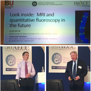
British Chiropractic Association Autumn Conference 2015, Bournemouth
I was delighted to have been invited to speak at the British Chiropractic Association’s Autumn Conference 2015, at the Marriot hotel, Bournemouth, perfectly situated to watch the sun glistening on the Channel. The beautiful weather was fitting for a particularly celebratory occasion with chiropractors travelling from all over the world to gather and celebrate 90 years of the BCA and 50 years of AECC. To be included as a speaker alongside some of the top researchers in the musculoskeletal field, and sharing the stage with no less than Professor Alan Breen, was slightly nerve-racking but also a very great honour.
Look Inside: MRI and quantitative fluoroscopy in the future
Alan Breen first started experimenting with what is now known as quantitative fluoroscopy (QF), taking measurements from motion x-rays, in the 1980s but it was not until the 2000s that computing power was sufficient to make this a reality. QF has since been found to be accurate and reliable in the measurement of inter-vertebral motion in both the cervical (my PhD work) and lumbar spines, and has been commercialised in the United States. It should start appearing in European hospitals within the next few years – watch this space. What will be possible with QF is a more accurate measurement of spinal stability to better inform surgical decisions and also the potential to better guide conservative treatment. The examples given from a case series during the presentation included showing that suspected lumbar instability is often not confirmed (therefore the potential to avoid unnecessary fusion surgery) and where it is present there is perhaps an increased chance of good surgical outcomes. Also, manipulation/mobilisation might be better targeted at identified segmental restrictions, and exercise therapy better directed where hypermobile or lax joint motion is present.
Alan’s part of the presentation included discussing what was possible with an open-MRI. Aside it being preferable for claustrophobic patients, the ability to image patients weight-bearing has meant the identification of disc hernia in some patients that would have been missed if imaged lying-down, therefore open-MRI is likely to play an important role in improving the diagnosis of persistent radicular pain. He also touched on diagnostic ultrasound which is showing value particularly in the diagnosis of soft-tissue injury of the extremities.
We are only at the beginning of finding out if the potential of QF (and other imaging techniques) will be fully realised, that of improved outcomes for patients with neck and back pain. Its use, like that of MRI, is likely to be restricted to those patients with particularly problematic spinal pain, but it (and open-MRI/diagnostic ultrasound) is a welcome addition to the diagnostic armamentarium of the chiropractor and other musculoskeletal professionals. And it was developed, not by a massive multinational corporation, but by a member of the BCA at AECC, a partner college of BU. Now that is worth celebrating.
Dr Jonathan (Jonny) Branney
Lecturer in Adult Nursing
Faculty of Health and Social Sciences
NB: This is a truncated version of a longer blog that can be found here


 REF Code of Practice consultation is open!
 BU Leads AI-Driven Work Package in EU Horizon SUSHEAS Project
 Evidence Synthesis Centre open at Kathmandu University
 Expand Your Impact: Collaboration and Networking Workshops for Researchers
 ECR Funding Open Call: Research Culture & Community Grant – Apply now
 ECR Funding Open Call: Research Culture & Community Grant – Application Deadline Friday 12 December
 MSCA Postdoctoral Fellowships 2025 Call
 ERC Advanced Grant 2025 Webinar
 Update on UKRO services
 European research project exploring use of ‘virtual twins’ to better manage metabolic associated fatty liver disease