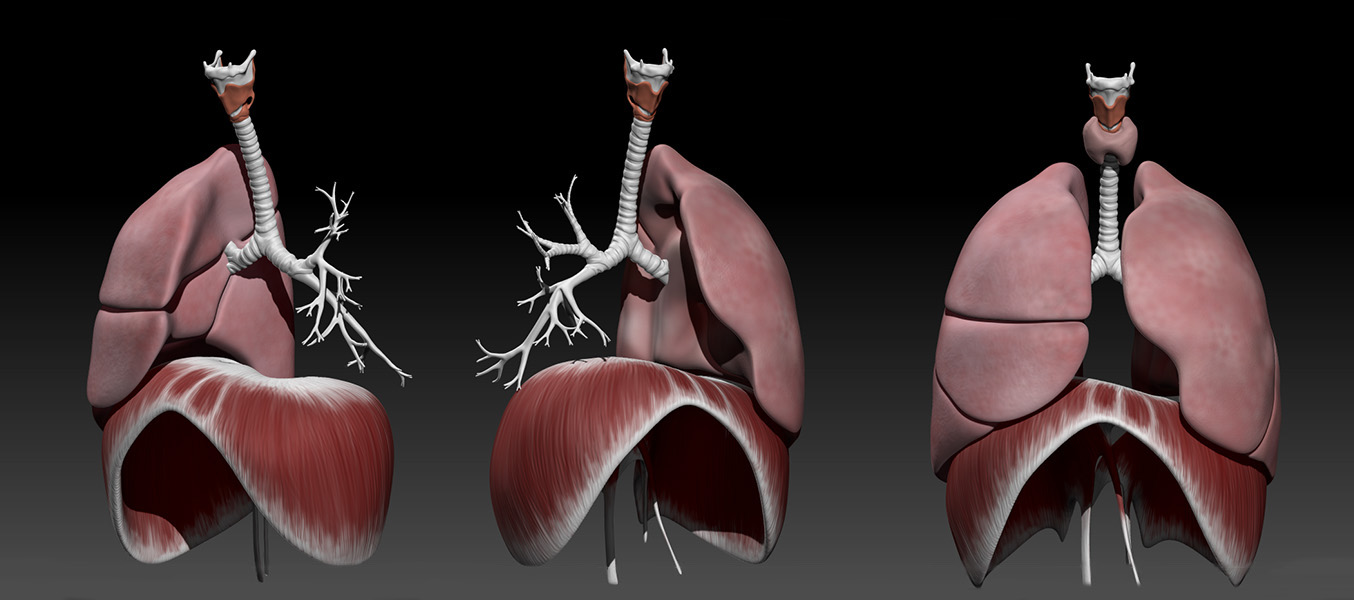
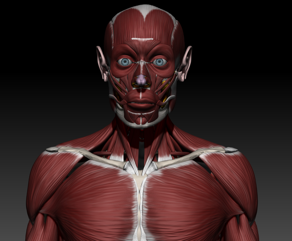
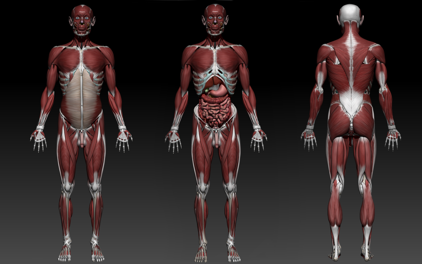
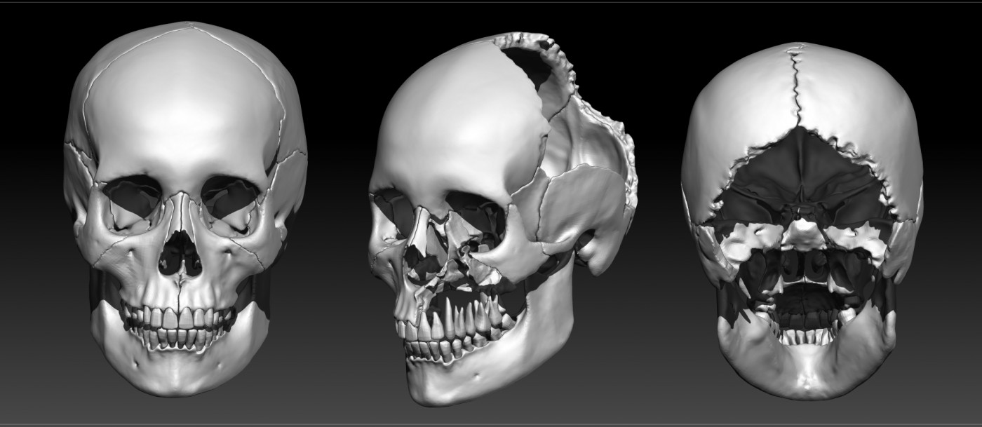
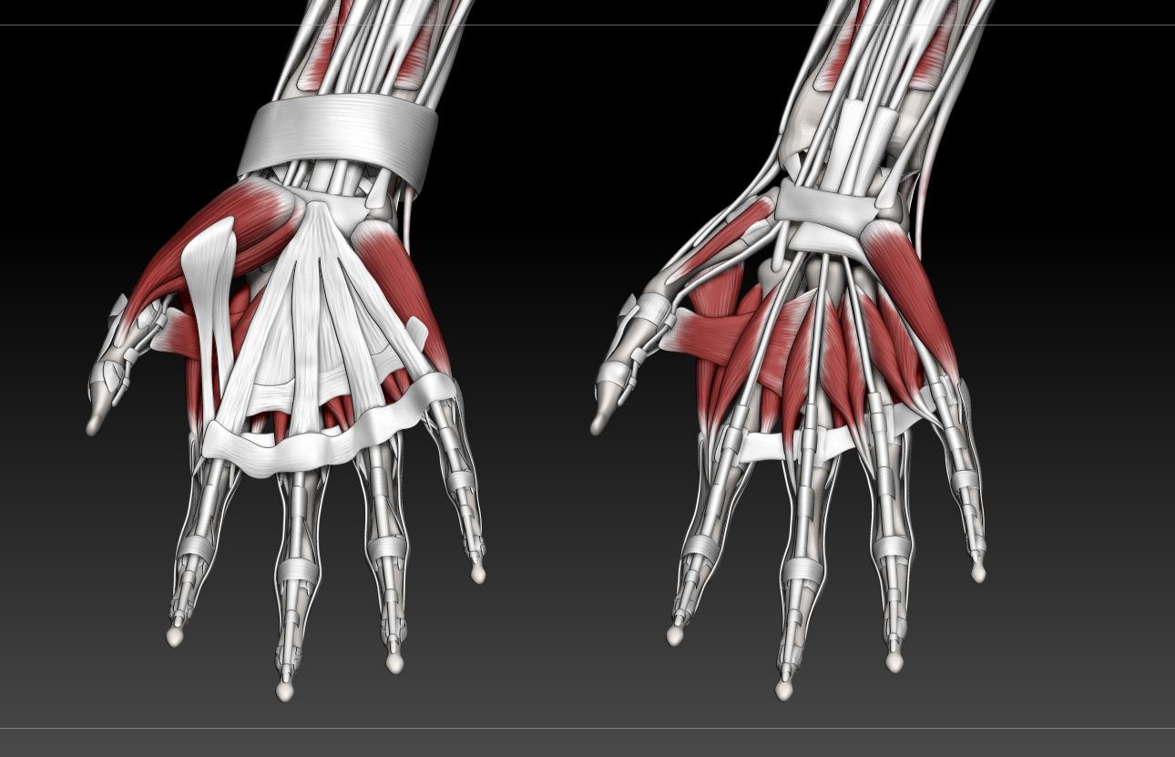
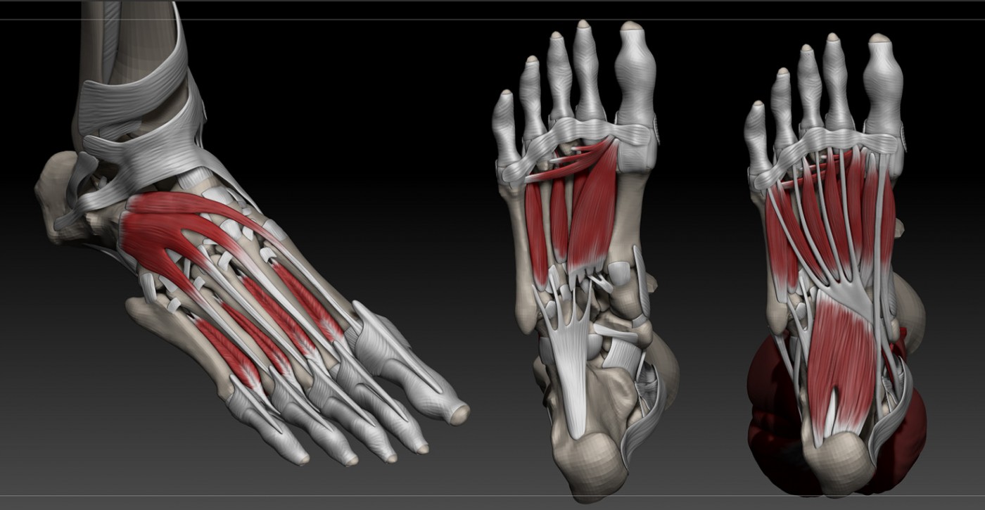
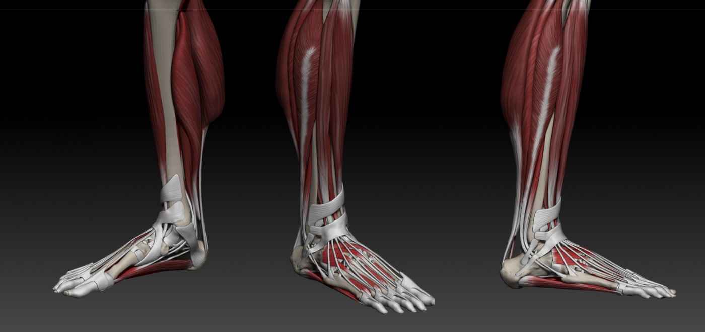
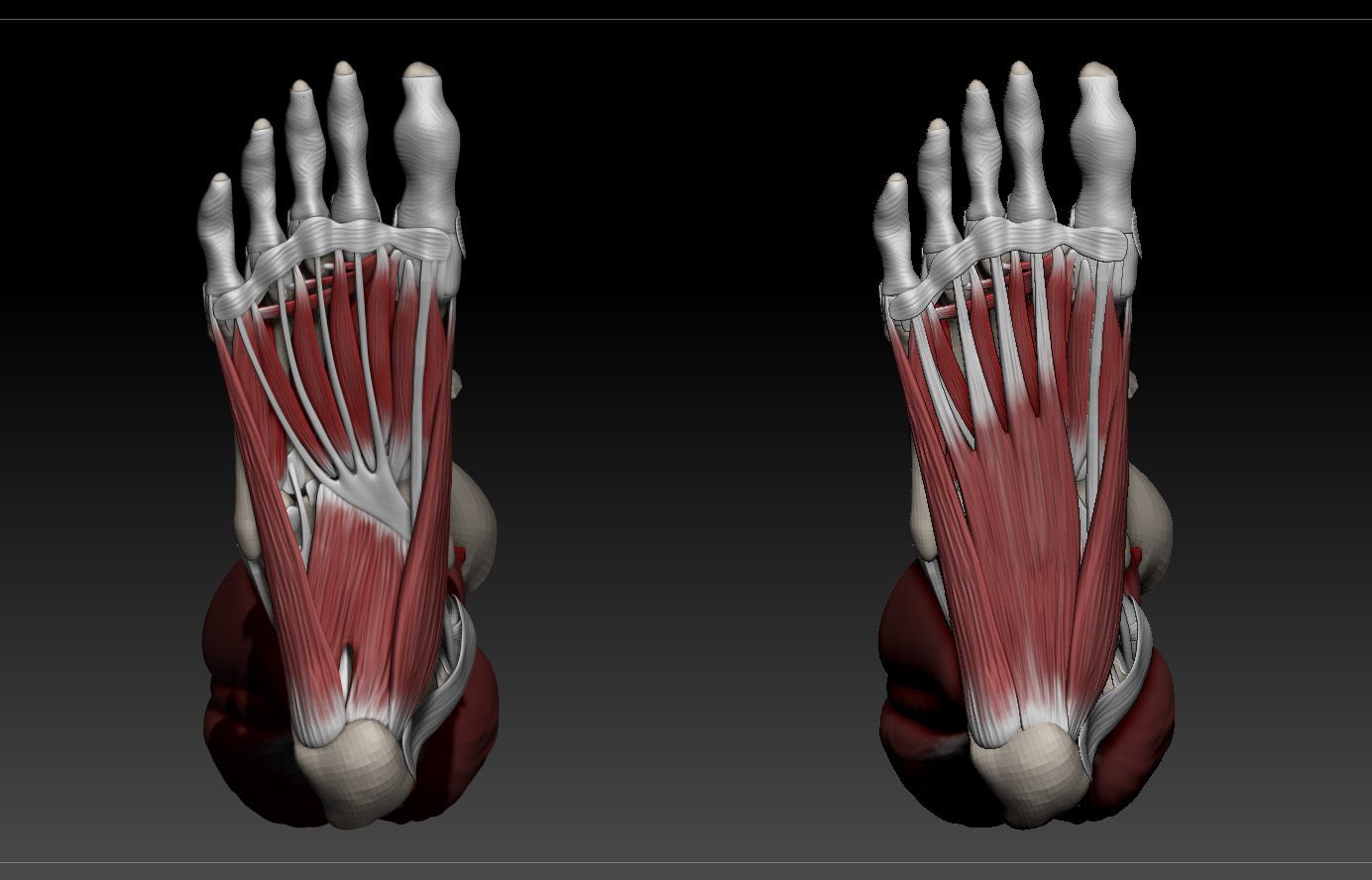
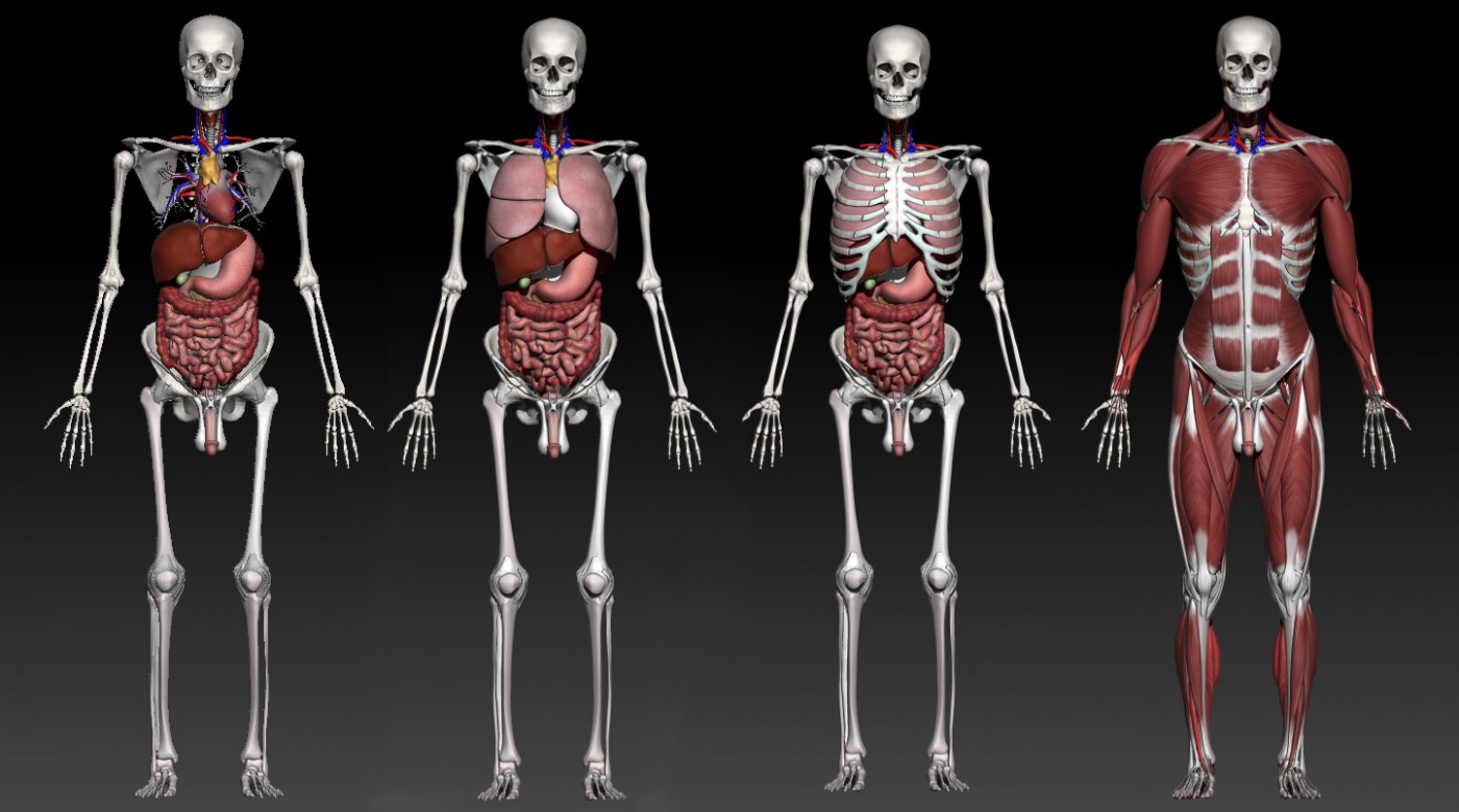
As one of the products from the HEIF6 Project, our team has developed a wide collection of digital assets to represent human anatomy. The understanding of human anatomy is vital to the delivery of healthcare. For medical students, this necessary awareness of anatomy and 3D spatial orientation is traditionally learned through cadaveric dissection. This is expensive and has practical as well as ethical constraints to available teaching time. The digital models can be used as assets for interdisciplinary research between the fields of Arts, Science and Healthcare. We welcome ideas from the BU community for proposals of novel use cases, research, grant applications and availability as teaching tools or base models for complex animation techniques.
Contact:
Learn more about the available assets and how to collaborate with the Neuravatar team by contacting Dr Xiaosong Yang (xyang@bournemouth.ac.uk) or Dr. Rupert Page (Rupert.Page@poole.nhs.uk).
👀 A glance at the 3D models available so far 👀

 Failing to publish data from clinical trials presents risk to human health


 3C Online Social: Thursday 26 March 1–2pm – Research Culture, Community & Can you Guess Who?
 INRC book roundtable/presentation by Drs Jonathan Cole and Catherine Talbot, Wednesday 22/04/2026, 13:00h, P426
 BU M.Res. student’s evidence to UK Parliamentary Women & Equalities Committee
 ECR Funding Open Call: Research Culture & Community Grant – Apply now
 ECR Funding Open Call: Research Culture & Community Grant – Application Deadline Friday 12 December
 MSCA Postdoctoral Fellowships 2025 Call
 ERC Advanced Grant 2025 Webinar
 Update on UKRO services
 European research project exploring use of ‘virtual twins’ to better manage metabolic associated fatty liver disease