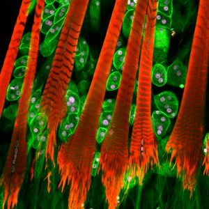
Dr Paul Hartley, Senior Lecturer in Functional Genetics, Faculty of Science & Technology
Honey Bee Heart
The first instalment of the returning ‘Photo of the Week’ series features Dr Paul Hartley’s image of a Honey Bee Heart. The series is a weekly instalment, which features an image taken by our fantastic BU staff and students. The photos give a glimpse into some of the fascinating work our researchers have been doing across BU and the wider community.
In this image we can see the pericardial muscles and oenocytes of a honey bee heart- these are stained red and green. The oenocytes act as toxin-treatment and excretion plants, to help maintain clean blood – much as our livers and kidneys do. The pericardial muscles hold the heart in place so that it can contract properly. Research has shown that human and insect cardiovascular systems share similar genetics. Dr Hartley’s research is based on a simple premise- if something causes disease in one organism, it probably causes disease and can be studied in the other. Dr Hartley took this picture using a Leica SP8 confocal microscope.
If you’d like to find out more about the research or the photo itself then please contact Dr Hartley.
This photo was orginially an entry to the 2017 Research Photography Competition. If you have any other questions about the Photo of the Week series or the competition please email research@bournemouth.ac.uk
 Photo of the Week: Pollen from a Bumblebee
 Photo of the Week: This is me. I am Ron.
 Photo of the Week: Zooming in on Dietary Differences
 Photo of the Week: Dramaturgical study of ‘Game of Thrones’


 Nursing Research REF Impact in Nepal
 Fourth INRC Symposium: From Clinical Applications to Neuro-Inspired Computation
 ESRC Festival of Social Science 2025 – Reflecting back and looking ahead to 2026
 3C Event: Research Culture, Community & Cookies – Tuesday 13 January 10-11am
 Dr. Chloe Casey on Sky News
 ECR Funding Open Call: Research Culture & Community Grant – Application Deadline Friday 12 December
 MSCA Postdoctoral Fellowships 2025 Call
 ERC Advanced Grant 2025 Webinar
 Horizon Europe Work Programme 2025 Published
 Update on UKRO services
 European research project exploring use of ‘virtual twins’ to better manage metabolic associated fatty liver disease